生命理工学系 News
【研究室紹介】 工藤研究室(~2017.3)
メダカを用いた骨の発生・形成メカニズムの解明
生命理工学系にはライフサイエンスとテクノロジーに関連した様々な研究室があり、基礎科学と工学分野の研究のみならず、医学や薬学、農学等、幅広い分野で最先端の研究が活発に展開されています。
研究室紹介シリーズでは、ひとつの研究室にスポットを当てて研究テーマや研究成果を紹介。今回は、多細胞組織の維持、再生の仕組みを解明する、工藤研究室です。
※工藤教授は2017年3月31日、東工大を退職いたしました。
生命理工学コース
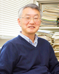
教授 工藤明![]()
| キーワード | メダカ、造骨細胞、破骨細胞 |
|---|
研究紹介
骨を作る造骨細胞と骨を壊す破骨細胞のバランスによって骨量が決まります。
このバランスが崩れると骨粗鬆症になります。
宇宙では無重力になり、骨量が減少することが知られています。
メダカを用いて生きたままこの造骨と破骨の細胞の分化、成熟、代謝を観察することにより、これまでマウスでは解明できなかった新しい骨の発生・形成メカニズムがわかります。

国際宇宙ステーションで飼育したダブルトランスジェニックメダカ
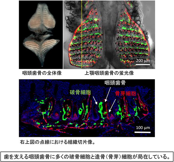
骨形成と骨吸収が盛んな咽頭歯骨
研究成果
- 代表論文
- [1] Chatani, M., Morimoto, H., Takeyama, K., Mantoku, A., Tanigawa, N., Kubota, K., Suzuki, H., Uchida, S., Tanigaki, F., Shirakawa, M., Gusev, O., Sychev, V., Takano, Y., Itoh, T. and Kudo, A. Acute transcriptional regulation in osteoblasts/osteoclasts immediately after exposure to microgravity, uncovered by cell imaging in medaka.Sci. Rep. 6: 39545 (2016)
- [2] Takeyama, K., Chatani, M., Inohaya, K. and Kudo, A. TGFβ-2 signaling is essential for osteoblast migration and differentiation during fracture healing in medaka fish. Bone 86: 68-78 (2016)
- [3] Mantoku, A., Chatani, M., Aono, K., Inohaya, K. and Kudo, A. Osteoblast and osteoclast behaviors in the turnover of attachment bones during medaka tooth replacement. Dev. Biol. 409: 370-381 (2016)
- [4] Chatani, M., Mantoku, A., Takeyama, K., Abduweli,D., Sugamori, Y., Aoki, K., Ohya, K., Suzuki, H., Uchida, S., Sakimura, T., Kono, Y., Tanigaki, F., Shirakawa, M., Takano, Y. and Kudo, A. Microgravity promotes osteoclast activity in medaka fish reared at the international space station. Sci. Rep. 5: 14172 (2015)
- [5] Taimatsu, K., Takubo, K., Maruyama, K., Suda, T. and Kudo, A. Proliferation following tetraploidization regulates the size and number of erythrocytes in the blood flow during medaka development, as revealed by the abnormal karyotype of erythrocytes in the medaka TFDP1 mutant. Dev. Dyn. 244: 651-668 (2015)
- [6] Takeyama, K., Chatani, M., Takano, Y. and Kudo, A. In-vivo imaging of the fracture healing in medaka revealed two types of osteoclasts before and after the callus formation by osteoblasts. Dev. Biol. 394: 292-304 (2014)
- [7] Ito, K., Morioka, M., Kimura, S., Tasaki, M., Inohaya, K. and Kudo, A. Differential reparative phenotypes between zebrafish and medaka after cardiac injury. Dev. Dyn. 243: 1106-1115 (2014)
- [8] Iida, Y., Hibiya, K., Inohaya, K. and Kudo, A. Eda/Edar signaling guides fin ray formation with preceding osteoblast differentiation, as revealed by analyses of the medaka all-fin less mutant afl. Dev. Dyn. Doi: 10.1002/DVDY.24120 (2014)
- [9] Fujita, M., Mitsuhashi, H., Isogai, S., Nakata, T., Kawakami, A., Nonaka, I., Noguchi, S., Hayashi, Y. K., Nishino, I. and Kudo, A. Filamin C plays an essential role in the maintenance of the structural integrity of cardiac and skeletal muscles, revealed by the medaka mutant zacro. Dev. Biol. 361: 79-89 (2012)
- [10] Chatani, M., Takano, Y. and Kudo, A. Osteoclasts in bone modeling, as revealed by in vivo imaging, are essential for organogenesis in fish. Dev. Biol. 360: 96-109 (2011)
- [11] Moriyama, A., Inohaya, K., Maruyama, K. and Kudo, A. Bef mutant reveals the essential role of c-myb in both primitive and definitive hematopoiesis. Dev. Biol. 345: 133-143 (2010)
- [12] Inohaya, K., Takano, Y. and Kudo, A. Production of wnt4b by floor plate cells is essential for the segmental patterning of the vertebral column in medaka. Development 137: 1807-1813 (2010)
- [13] Ohisa, S., Inohaya, K., Takano, Y. and Kudo, A. sec24d encoding a component of COPII is essential for vertebra formation, revealed by the analysis of the medaka mutant, vbi. Dev. Biol. 342: 85-95 (2010)
- [14] Hibiya, K., Katsumoto, T., Kondo, T., Kitabayashi, I. and Kudo, A. Brpf1, a subunit of the MOZ histone acetyl transferase complex, maintains expression of anterior and posterior Hox genes for proper patterning of craniofacial and caudal skeletons. Dev. Biol. 329: 176-190 (2009)
教員紹介
工藤明 教授 薬学博士
| 1977年 3月 | 東京工業大学 総合理工学研究科 修士修了 |
|---|---|
| 1994年 12月より | 現職 |
教員からのメッセージ
- 工藤教授より
-
メダカ、ゼブラフィッシュが代表する小型魚類は、器官形成のメカニズムを解明するのに最も注目されている実験動物です。
iPS細胞を始めとする幹細胞学が盛んですが、次の科学は、細胞から心臓、筋肉、骨などの器官がどのようにしてできるかに焦点が注がれています。
- 研究室と研究テーマ
- 無重力で骨関連遺伝子以外でも発現が急上昇する遺伝子を発見|生命理工学系 News
- 無重力による骨量減少メカニズムの一端を解明|東工大ニュース
- メダカ飛行士、再び宇宙へ|東工大ニュース
※この内容は掲載日時点の情報です。
