Life Science and Technology News
Novel nanoparticle MRI probe enables quantitative pH imaging
Researchers have developed a contrast agent that distinguishes subtle acidity differences between cancerous and normal tissues
Accurately measuring tissue acidity could improve disease diagnosis and treatment monitoring. Researchers from Science Tokyo and collaborating institutions have developed a nanoparticle contrast agent that enables quantitative pH imaging using magnetic resonance imaging. The probe designed by tuning the crosslinked polymer shell distinguishes pH values from 6.0 to 7.5 independently of its concentration, offering a promising approach for investigating tumor acidity, assessing disease progression, and monitoring treatment responses.
Nanoparticle MRI Probe-Enabled Quantitative pH imaging
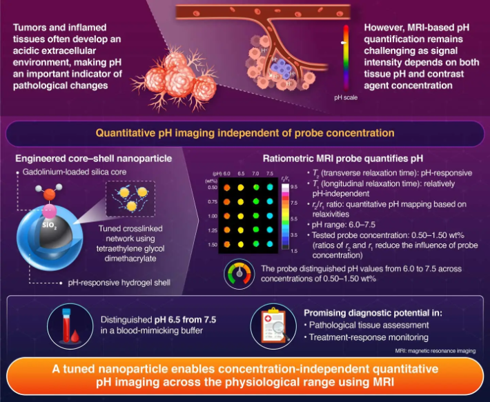
The acidity of tissues can provide important clues about a disease. Tumors and inflamed tissues often become more
acidic than their surroundings because of changes in cellular metabolism. Measuring these pH variations could help
researchers understand disease progression, identify abnormal tissue environments, and evaluate treatment responses.
However, detecting small pH differences inside the body remains challenging because magnetic resonance imaging (MRI)
signals are affected by the concentration of the imaging agent used.
MRI provides a way to visualize deep tissues without exposing the body to ionizing radiation. To visualize tissue
acidity, scientists have developed pH-responsive MRI contrast agents, which alter the MRI signal in response to
changes in pH. However, the signal produced by these agents also depends on how much of the agent (probe) is present
in the tissue. Consequently, a stronger or weaker signal does not necessarily indicate a change in acidity,
complicating accurate pH measurement.
To address this limitation, a research team led by Associate Professor Satoshi Okada and Professor Hiroyuki Nakamura
of the Laboratory for Chemistry and Life Science, Institute of Science Tokyo (Science Tokyo), Japan, has developed a
new nanoparticle contrast agent for quantitative pH imaging within the body using MRI. The research was conducted in
collaboration with Specially Appointed Assistant Professor Ken Kato and Professor Koichi Kato of the National
Institutes of Natural Sciences, Japan, and Professor Kazuya Kikuchi of the University of Osaka, Japan.
The study, published online in the journal Small on September 23, 2026, presents a
strategy for improving MRI-based pH measurements by precisely tuning the structure of a pH-responsive polymer
network.
The team developed a core–shell nanoparticle comprising a silica core containing gadolinium-based MRI contrast
agents and an outer shell made of a pH-responsive polymer hydrogel. They modified the shell's crosslinked network
using tetraethylene glycol dimethacrylate, a chemical crosslinker that helps connect polymer chains while providing
flexibility to the network. Okada explains about the core–shell nanoparticle, “As the surrounding environment became
more acidic, the polymer shell shrinks, changing the nanoparticle's interaction with surrounding water and its
transverse relaxation behavior. Meanwhile, its longitudinal relaxation response remained largely unchanged by
pH.”
To quantify these changes, the researchers employed ratiometric MRI, an approach that compares two
relaxation-related measurements in order to reduce the influence of contrast agent concentration. They calculated
the ratio of transverse to longitudinal relaxivity (r2/r1), allowing pH to be
estimated without relying solely on MRI signal intensity. The nanoparticle successfully distinguished pH values of
6.0, 6.5, 7.0, and 7.5 across contrast agent concentrations ranging from 0.50 to 1.50 wt%. The probe also
distinguished between pH 6.5 and 7.5 in the presence of fetal bovine serum, a biological material used to evaluate
performance under laboratory conditions that approximates the aspects of biological environments.
The study findings highlight how modifying the molecular structure of a polymer shell can improve the sensitivity
and reliability of quantitative MRI. By reducing the influence of contrast agent concentration, the technique could
aid in distinguishing acidity differences in tissues more accurately. Okada explains, “Our study supports the
potential for new MRI-based diagnostic approaches to accurately assess cancer progression and disease status, as
well as for methods to monitor treatment response.”
In the future, incorporating targeting molecules to enhance probe accumulation in specific tissues could contribute
to more reliable pH imaging, facilitating the investigation of disease-associated pH changes and potentially
improving the assessment of cancer progression and treatment response.
- Reference
| Authors: | Ken Kato1,2,3, Akira Sumiyoshi4, Kyoko Katsumata5, Hiroyuki Nakamura5, Koichi Kato1,2,3,6, Kazuya Kikuchi1,2,7*, Satoshi Okada5* |
|---|---|
| Title: | A Ratiometric MRI Probe with a Tuned Crosslinked Network for Quantitative Imaging of Physiological pH |
| Journal: | Small |
| DOI: | 10.1002/smll.75796 |
| Affiliations: | 1Core for Spin Life Sciences, National Institutes of
Natural Sciences, Japan 2Exploratory Research Center on Life and Living Systems (ExCELLS), National Institutes of Natural Sciences, Japan 3Institute for Molecular Science (IMS), National Institutes of Natural Sciences, Japan 4Institute for Quantum Medical Science, National Institutes for Quantum Science and Technology (QST), Japan 5Laboratory for Chemistry and Life Science, Institute of Science Tokyo, Japan 6Graduate School of Pharmaceutical Sciences, Nagoya City University, Japan 7Department of Applied Chemistry, The University of Osaka, Japan |
*Corresponding authors
Related articles
- Satoshi Okada | Science Tokyo Research Information DB - Science and engineering fields
- Hiroyuki Nakamura | Science Tokyo Research Information DB - Science and engineering fields
- Nakamura & Okada Laboratory
- Laboratory for Chemistry and Life Science
- Department of Life Science and Technology, School of Life Science and Technology
- Institute of Integrated Research
- School of Life Science and Technology
- Total Health Design Visionary Initiative
Further information
Associate Professor Satoshi Okada
Institute of Integrated Research, Institute of Science Tokyo
Email okada@cls.iir.isct.ac.jp
Featured News