Life Science and Technology News
【Labs spotlight】 Kondoh Laboratory(until Mar. 2023)
Beating hypoxic cancer by novel technology & ideas
The Department has a variety of laboratories for Life Science and Technology, in which cutting-edge innovative research is being undertaken not only in basic science and engineering but also in the areas of medicine, pharmacy, agriculture, and multidisciplinary sciences.
This "Spotlight" series features a laboratory from the Department and introduces you to the laboratory's research projects and outcomes. This time we focus on Kondoh Laboratory.
※Professor Kondoh was retired on March 31, 2023, as she reached the mandatory retirement age.

Human Centered Science and Biomedical Engineering Graduate Major
Professor Shinae Kondoh![]()
| Degree | PhD (D.M.Sc) 1989, Osaka University |
|---|---|
| Areas of Research | Molecular Oncology, in vivo imaging, Cancer therapy. |
| Keywords | Cancer biology, Protein engineering, Optical imaging, Drug development |
| Website | Kondoh Laboratory |
Research interest
Nowadays, one of two people experiences cancer in life. Long-term cure of the diseases is still challenging even with the most advanced therapy. In Kondoh laboratory, we have brought advanced biotechnologies in cancer research field to develop innovative therapeutic strategies based on medicine-engineering collaboration.
- Development of anti-cancer drugs targeting tumor microenvironment
- Tumor-specific environment (tumor microenvironment) is a promising target for cancer therapy. For example, in most types of solid tumors, uncontrolled tumor growth and immature blood vessels during angiogenesis often generate hypoxic microenvironment (Fig.1). Under hypoxic conditions, hypoxia-inducible factors are activated, inducing a vast array of gene products that promote both tumor progression and resistant to therapies. Thus, tumor hypoxia can be novel "microenvironmental target" for cancer therapy. We have addressed a novel strategy to target hypoxic cancer cells by using fusion protein drugs (TOP3, POP33) that selectively kill the HIF-active cancer cells. We currently optimize treatment protocol for combination therapy and preparation of fusion protein drugs for clinical test on patients with pancreatic cancers.
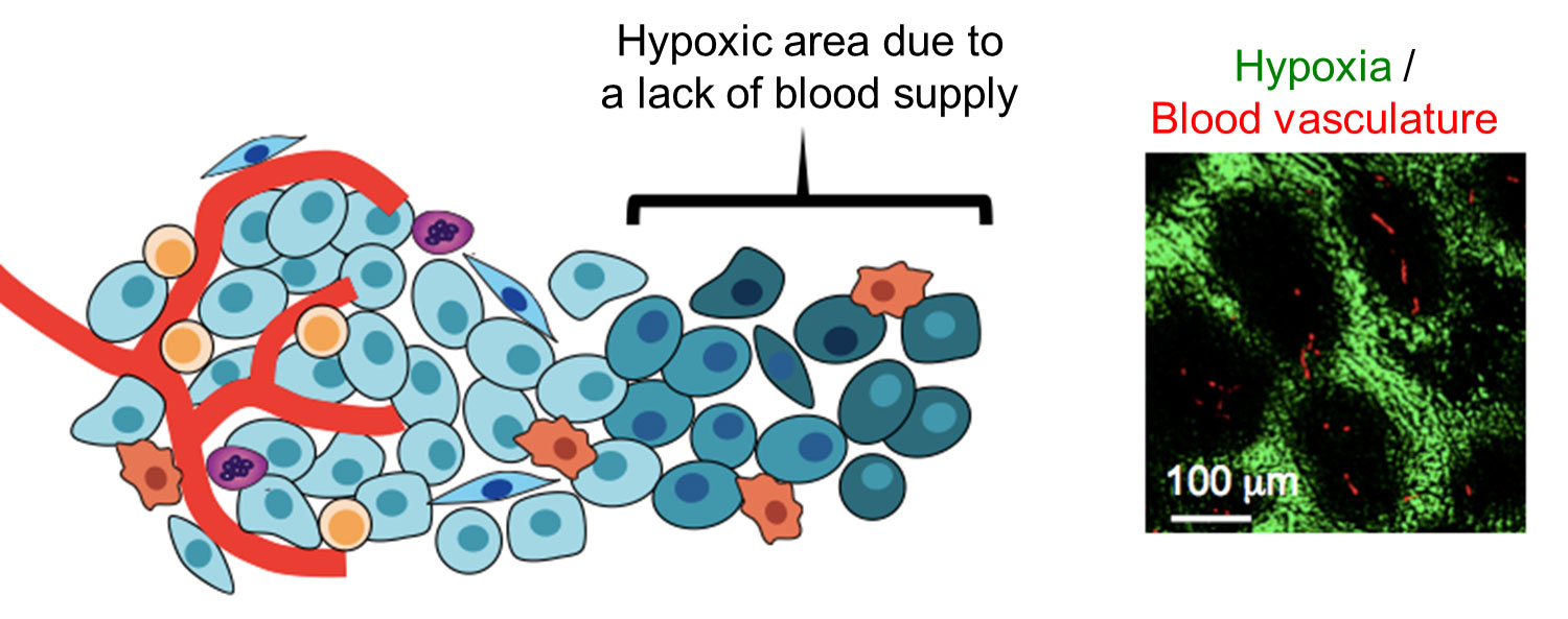
Fig1. Hypoxic microenvironment in tumor tissues
- Development of peptide drugs as an antibody drug alternative
- Small proteins/peptides that have a high affinity for cancer cell surface markers (antibody mimetics) are promising low-cost alternatives to antibody. Therefore, various types of antibody mimetics have been extensively developed. We recently found that peptides structurally constrained in a protein scaffold were able to bind to their target molecule more strongly and specifically than the fluctuating them. Currently we are optimizing the scaffolds in order to increase binding activity of antibody mimetics to the levels comparable to antibody.
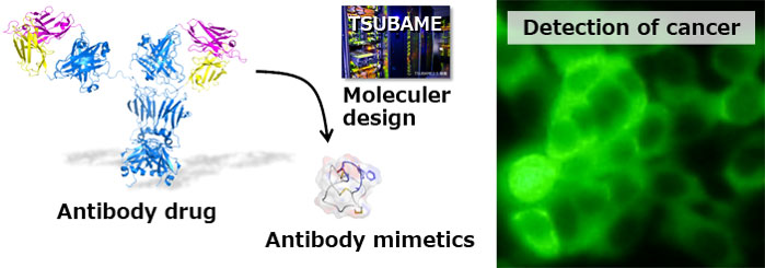
Fig2. Detection of cancer cells using antibody mimetics
- Next-generation drug discovery targeting treatment-resistant cancer cells
- The drug-resistant and/or dormant cancer cells often survive after treatments and become a hotbed for recurrence. Therefore, it is important to target these malignant cancer cells for complete recovery but there are fewer drugs targeting these cells. We thus have been developing next-generation drugs through identification of novel immature myeloid cells which significantly promoted the growth of tumor, drug screening targeting inducible-dormant cancer cells, and development of drug delivery system (DDS) using cell penetrating peptide (CPP).

Fig3. Accumulation of CPP-fused probe into tumor
Development of optical imaging tools illuminating tumor microenvironment
It is increasingly recognized that tumor-specific microenvironment critically direct tumor malignant processes. Understanding of microenvironmental regulation of tumor malignancy would pave the way to develop novel therapeutic strategies. However, traditional "static" biological assays are not sufficient to precisely dissect the mechanisms regulated by "dynamic" tumor microenvironment (TME). We thus have developed imaging tools to noninvasively monitor TME such as intratumoral hypoxia. We currently work on development of new bioluminescence imaging tools that allow for multifaceted approaches to understand tumor malignancy process regulated by TME.

Fig4. Development of optical imaging tools for visualizing TME
Understanding of molecular mechanisms regulating metastasis
Metastasis remains a major cause of death from solid tumors. Understanding of critical mechanisms regulating metastasis holds a promise to improve prognosis of patients and overcome cancer diseases in the future. Metastasis is a highly complex process including multi-step cascade. To fully understand this process, interactions between cancer cells and stromal environments are critically important as well as genetic characterizations of cancer cells. To address this issue, we have combined traditional biological approaches with noninvasive imaging, unique animal models and bioinformatics. In particular, we currently study on lung metastasis of osteosarcoma and bone metastasis of prostate cancers.

Fig5. Interactions between cancer cells and tissue stromal cells in a metastatic process
Research findings
Selected publications
- [1] Kadonosono T, Yimchuen W, Tsubaki T, Shiozawa T, Suzuki Y, Kuchimaru T, Sato Y, Kizaka-Kondoh S. Domain architecture of vasohibins required for their chaperone-dependent unconventional extracellular release. Protein Sci, in press.
- [2] Kuchimaru T, Suka T, Hirota K, Kadonosono T, Kizaka-Kondoh S. A novel injectable BRET-based in vivo imaging probe for detecting the activity of hypoxia-inducible factor regulated by the ubiquitin-proteasome system. Sci Rep 6, 34331 (2016).
- [3] Kuchimaru T, Iwano S, Kiyama M, Mitsumata S, Kadonodono T, Niwa H, Maki S, Kizaka-Kondoh S. A luciferin analogue generating near-infrared bioluminescence achieves highly sensitive deep-tissue imaging. Nat Commun 7, 11856 (2016).
- [4] Hoang N, Kadonosono T, Kuchimaru T, Kizaka-Kondoh S. HIF-targeting prodrug TOP3 combined with gemcitabine or TS-1 improves pancreatic cancer survival in an orthotopic model. Cancer Sci, 107, 1151-1158 (2016).
- [5] Kadonosono T, Yamano A, Goto T, Tsubaki T, Niibori M, Kuchimaru T, Kizaka-Kondoh S. Cell penetrating peptides improve tumor delivery of cargos through neuropilin-1-dependent extravasation. J Controlled Release 201, 14-21 (2015).
- [6] Kuchimaru T, Hoshino T, Aikawa T, Yasuda H, Kobayashi T, Kadonosono T, Kizaka-Kondoh S. Bone resorption facilitates osteoblastic bone metastatic colonization by cooperation of insulin-like growth factor and hypoxia. Cancer Sci 105, 553-559 (2014).
- [7] Ueda M, Ogawa K, Miyano A, Ono M, Kizaka-Kondoh S, Saji H. Development of an Oxygen-Sensitive Degradable Peptide Probe for the Imaging of Hypoxia-Inducible Factor-1-Active Regions in Tumors. Mol Imaging Biol 15(6): 713-721 (2013).
- [8] Kadonosono T, Kuchimaru T, Yamada S, Takahashi Y, Murakami A, Watanabe H, Tani T, Inoue M, Tsukamoto T, Toyoda T, Tanaka T, Hirota K, Urano K, Machida K, Eto T, Ogura T, Tsutsumi H, Ito M, Hiraoka M, Kondoh G & Kizaka-Kondoh S. Detection of the onset of ischemia and carcinogenesis by hypoxia-inducible transcription factor-based in vivo bioluminescence imaging. PLoS ONE 6(11):e26640 (2011).
- [9] Kuchimaru T, Kadonosono T, Tanaka S, Ushiki T, Hiraoka M, Kizaka-Kondoh S. In vivo imaging of HIF-active tumors by an oxygen-dependent degradation protein probe with an interchangeable labeling system. PLoS ONE 5(12): e15736 (2010).
- [10] Kizaka-Kondoh S, ltasaka S, Zeng L, Tanaka S.Zhao T. Takahashi Y, Shibuya K, Hirota K, Semenza GL, Hiraoka M. Selective killing of hypoxia-inducible factor-1-active cell improves survival in a mouse model of invasive and metastatic pancreatic cancer. Clin Cancer Res 15(10): 3433-3441 (2009).


