Life Science and Technology News
Shedding New Light: A New Type of Immunosensor for Immunoassay Tests
Immunoassays have emerged as an immensely reliable means of detection in fields ranging from biological research to food safety management. In a recent study, scientists at Tokyo Institute of Technology (Tokyo Tech) developed a new type of immunosensor for these purposes. Termed "BRET Q-body", the sensor works on the principle of bioluminescence resonance energy transfer (BRET) and can be used to detect antigens without the need for an external light source, greatly simplifying immunoassays.
Immunosensors are widely used in immunoassays to detect antigens. One such immunosensor is a quenchbody (Q-body), which contains a modified antibody fragment with a quenched fluorescent dye. When an antigen binds to the Q-body, the dye leaves the antibody and the fluorescence intensifies. The change in fluorescence intensity is easy to measure, making Q-body-based antigen detection systems incredibly simple. However, this method requires an external light source to excite the electrons in the fluorescent dye to produce luminescence.
One way to solve this is to induce luminescence by an alternative method. To achieve this, researchers from Tokyo Tech, Japan, have developed a novel immunosensor. They used a modified luciferase enzyme called "NanoLuc" (Nluc) that is originally responsible for bioluminescence in shrimp and fused it to the Q-body. This immunosensor, termed "BRET Q-body", works on the bioluminescence resonance energy transfer (BRET) principle. Here, a luminescent substrate is added to the fused Q-body. The substrate reacts with the enzyme and this reaction provides the energy required by the dye to induce fluorescence. This type of luminescence is advantageous, as Professor Hiroshi Ueda, who leads the team of researchers, explains, "The BRET Q-body system can be used to visualize the presence or the absence of an antigen as a change in the emission color without any instrument." Their findings were recently published in the journal Analytical Chemistry![]()
To prepare the BRET Q-body, the researchers used a single-chain antibody fragment which binds to the antigen BGP, a protein found in the bone. The antibody fragment was then labeled with a fluorescent dye. They then tested the fluorescence intensity of the resulting BRET Q-bodies and observed that the addition of the antigen increased the intensity of the fluorescence.
After these initial results, the researchers tested the antigen dependency of the BRET Q-body. They compared the fluorescence obtained by the new BRET-based method against the conventional irradiation-based method. The fluorescence intensity of the BRET-Q body was initially measured with excitation light and later in the presence of the luminescent substrate. They found that the antigen-binding brought the Nluc enzyme and the dye closer together resulting in higher fluorescence intensity levels when the substrate was used. As luminescence from the BRET Q-body is obtained initially from the enzyme and then from the fluorescent dye upon antigen binding, a simple color change indicates the presence of an antigen.
This study paves the way forward for a new class of bioluminescent sensors that do not require an external excitation light source. The novel BRET Q-bodies are expected to make immunoassay tests much simpler and more accurate. As Prof. Ueda explains the advantages and potential applications of their findings, "The detection of the BRET signal does not need a light source, allows visual observation of the color change, and easier integration to a smartphone-based device. Therefore, we expect that BRET Q-bodies will be a promising tool for diagnosis, food safety, environmental preservation, and biological research."
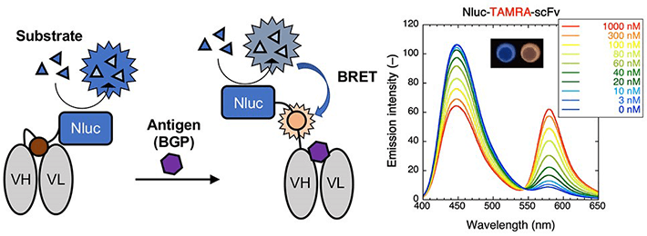
Figure 1.Mechanism for luminescence and emission spectra for the TAMRA-labeled BRET Q-body
The enzyme "Nluc" is added to the Q-body forming a Bret Q-body. Luminescence is observed upon antigen binding. To the right, emission spectra in the presence of the substrate of the BRET-Q body labeled with the fluorescent dye TAMRA-C5-mal can be observed. The inset shows the change in color observed on antigen binding.
- Reference
| Authors : | Riho Takahashi$, Takanobu Yasuda$, Yuki Ohmuro-Matsuyama and Hiroshi Ueda* |
|---|---|
| Title of original paper : | BRET Q-Body: A Ratiometric Quench-based Bioluminescent Immunosensor Made of Luciferase?Dye?Antibody Fusion with Enhanced Response |
| Journal : | Analytical Chemistry |
| DOI : | |
| Affiliations : | Tokyo Institute of Technology. |
| $Equal contributions * Corresponding author's email: |
- Biosensor for detecting proteins made entirely of a protein | Tokyo Tech News
- Ueda-Kitaguchi Lab.
- Researcher Profile | Tokyo Tech STAR Search - Hiroshi Ueda
- Laboratory for Chemistry and Life Science, Institute of Innovative Research
- Institute of Innovative Research (IIR)
- Department of Materials Science and Engineering, School of Materials and Chemical Technology
- Latest Research News
Further Information
Professor Hiroshi Ueda
Institute of Innovative Research, Tokyo Institute of Technology
E-mail : ueda@res.titech.ac.jp
Featured News


![]()