Life Science and Technology News
Shedding light on phototactic behavior of green algae
Scientists at Tokyo Institute of Technology and colleagues report that optical shielding by the eyespot pigments in Chlamydomonas reinhardtii, a unicellular green alga, determines the direction of their movement relative to light sources, and that the cell body behaves as a convex lens to focus and condense incident light.
Chlamydomonas reinhardtii (Chlamy) is a unicellular green alga that lives in fresh water throughout the world, and notably, the cells can change their swimming directions upon sensing light?referred to as phototactic behavior. However, in spite of their widespread use as a model organism, the role of pigments in the subcellular organelle called 'eyespot' in determination of the sign of phototaxis (direction of cell movement relative to light sources) is still unclear. Now, Ken-ichi Wakabayashi, who teaches Human Centered Science and Biomedical Engineering at Tokyo Tech and collaborators report in PNAS journal, "that the light shielding property of the eyespot pigments is essential for determination of phototactic sign."
Wakabayashi and coworkers observed a reversal in the sign of phototaxis in newly isolated Chlamy mutant cells with missing eyespot pigments. To explain these findings, the researchers designed experiments to determine whether the Chlamy cells behaved as lenses to focus and condense light onto photoreceptors that are located on the "equatorial region" of the surface of one side of Chlamy [Fig.1].
Specifically, in normal 'wild type' cells the eyespot pigment is located below the photoreceptors, so when the eyespot is facing a light source, the light signal is reflected and amplified by the pigment layers constituting the eyespot onto the photoreceptors. On the other hand, when the eyespot and photoreceptors are pointing in the opposite direction to the light source, the light does not reach the photoreceptors because it is shielded by the eyespot pigments, and the cell does not change its swimming direction.
Furthermore, in the case of mutant cells without eyespot pigments, when the photoreceptors are pointing directly towards a light source, the light signal is incident directly onto the photoreceptors but it is too weak to trigger turning of the cells. In the opposite orientation, with the photoreceptors are pointing away from the light source, the light signal is focused and condensed by the lens-action of the cell onto the photoreceptors, leading to the turning of Chlamy.
The researchers state that, "isolation of the new mutant, lts1-211, revealed that the cellular lens effect affects cellular behavior in the absence of the eyespot pigments. The eyespot pigments therefore have a crucial role in determining the sign of phototaxis, by shielding the photoreceptors from light condensed by the cellular lens onto the back of the eyespot."
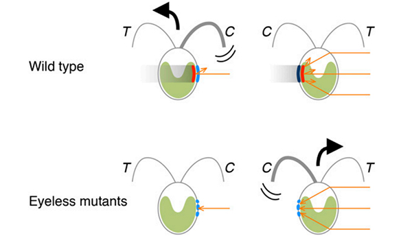
Figure 1. Illustration showing movements in opposite directions triggered by the relative orientation of the light source, eyespot pigments, and photoreceptors.
Background
Chlamy exhibits phototactic behavior and are widely used by scientists to model the functions of cells, such as cell movements and response to light for photosynthesis. It is well known that Chlamy moves in their aqueous environments by beating flagella, a hair-like organelle that generate force for swimming. Furthermore, Chlamy is phototactic, that is, they swim and find locations in their environments for optimizing photosynthesis by either moving towards light (positive phototaxis) or away from it (negative phototaxis). However, the role of the pigment (carotenoid) layers of the eyespot in determining the sign of phototaxis in Chlamy is not fully understood.
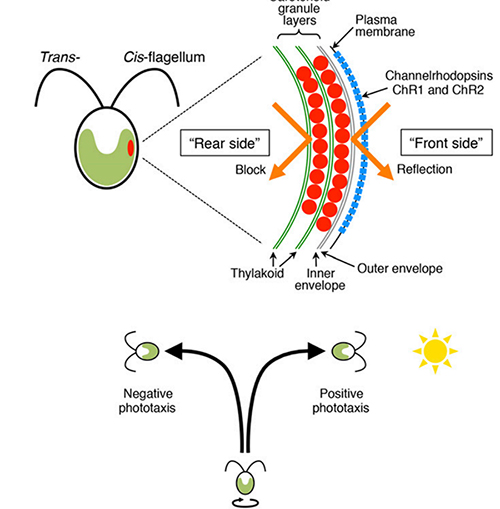
Figure 2. Schematics of cross section of Chlamydomonas cell and phototactic behavior relative to a light source.
Experimental details
Isolation of eyespot-less Chlamydomonas mutants with a reversed sign of phototaxis
The mutant lts1-211 that exhibits opposite phototactic sign compared to wild type was isolated by treatment with redox reagents against randomly mutagenized mutant pools; it shows positive phototaxis after treatment with reactive oxygen species (ROS) scavengers and negative phototaxis with ROS treatment. Importantly, most lts1-211 cells did not have a detectable eyespot.
Confirmation of the lens effect in green Chlamydomonas cells
The existence of focusing due to a lens effect in green Chlamydomonas cells was confirmed by observing both wild-type and lts-211 cells under a microscope with sideways illumination. A small bright area was observed on the side of the cell edge opposite to the light source, irrespective of the location of the eyespot. Furthermore, image formation by the cellular lens effect was also observed.
Significance
The methodology and findings of this research are expected to find applications in a wide range of research in molecular biology including the determination of the "refractive indices of spheroidal cellular organisms without special equipment."
Reference
| Authors: | Noriko Uekia,b, Takahiro Idea, Shota Mochijic, Yuki Kobayashia, Ryutaro Tokutsud,e,f, Norikazu Ohnishid, Katsushi Yamaguchig, Shuji Shigenobue,g, Kan Tanakaa,f, Jun Minagawad,e,f, Toru Hisaboria,f, Masafumi Hironoh and Ken-ichi Wakabayashia |
|---|---|
| Title of original paper: | Eyespot-dependent determination of the phototactic sign in Chlamydomonas reinhardtii |
| Journal: | Proceedings of the National Academy of Sciences of the United States of America (PNAS) |
| DOI : | 10.1073/pnas.1525538113 |
| Affiliations: | aLaboratory for Chemistry and Life Science, Institute of Innovative Research, Tokyo Institute of Technology bDepartment of Biological Sciences, Graduate School of Science and Engineering, Chuo University cDepartment of Biological Sciences, Graduate School of Science, University of Tokyo dDivision of Environmental Photobiology, National Institute for Basic Biology eDepartment of Basic Biology, Faculty of Life Science, The Graduate University for Advanced Studies fCore Research for Evolutional Science and Technology, Japan Science and Technology Agency gFunctional Genomics Facility, National Institute for Basic Biology hDepartment of Frontier Bioscience, Hosei University |
Further information
Associate Professor Ken-ichi Wakabayashi
Laboratory for Chemistry and Life Science,
Institute of Innovative Research
Email wakaba@res.titech.ac.jp
Tel +81-45-924-5235
Featured News


![]()