Life Science and Technology News
Illuminating detection of deep cancers
Tokyo Tech and UEC researchers develop a new bioluminescence imaging system to improve detection sensitivity of targets in deep tissues
To see right through something is to know its true intent—the same could be said to apply to biological tissues, especially tumors.
Bioluminescence imaging with a firefly enzyme, called luciferase, and its substrate D-luciferin, is widely used to monitor biological processes. However, the emission wavelength of bioluminescence produced by D-luciferin limits the sensitivity of this technique. At 562 nm, this light does not effectively penetrate biological tissues.
To overcome this limitation, a team of Tokyo Tech and the University of Electro-Communications (UEC) researchers developed a luciferin analog (a compound that resembles another in structure) that can produce bioluminescence with near-infrared wavelength and is applicable in animal experiments. This allows markedly higher target-detection sensitivity, even at very low concentrations.
A novel soluble luciferin analog
The UEC researchers had previously synthesized a novel luciferin analog, AkaLumine, by altering the chemical structure of D-luciferin. While the emission wavelength of bioluminescence produced by AkaLumine yielded high penetration, its insolubility hindered its use. The team moved beyond this to screen for water-soluble derivatives of AkaLumine, and discovered that one of them, AkaLumine hydrochloride (AkaLumine-HCl), was in fact soluble. The Tokyo Tech researchers evaluated these substrates and had provided proper information for directing it to be practical use in animal experiments, making AkaLumine-HCL applicable for bioluminescence imaging of deep tissues.
AkaLumine-HCl emitted near-infrared bioluminescence at 677 nm when reacted with firefly luciferase (Figure 1), and had greatly improved tissue-penetration efficiency. In 4-mm or 8-mm slice of beef, AkaLumine-HCl bioluminescence showed penetration 5-fold and 8.3-fold higher than bioluminescence produced by D-Luciferin (Figure 2). Notably, achieving such a high sensitivity using D-luciferin would require a 60-fold higher concentration.
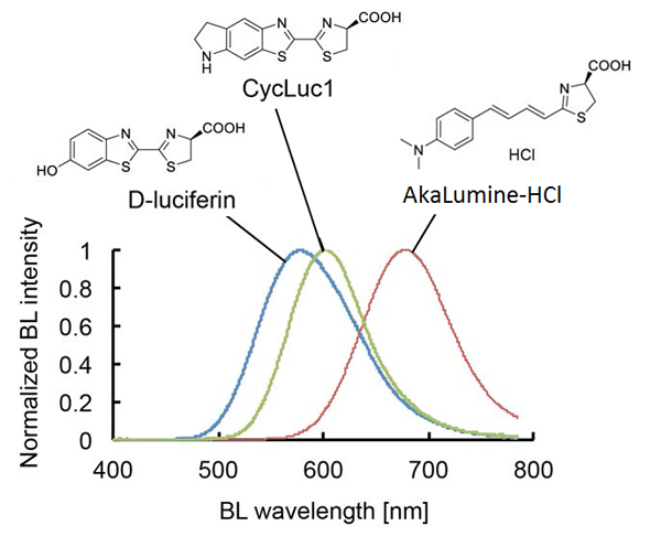
Figure 1. Bioluminescence emission spectra of D-luciferin, CycLuc1 or AkaLumine-HCl.
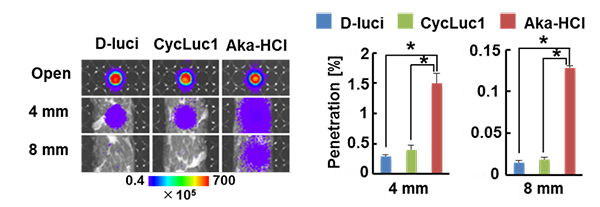
Figure 2. Biological tissue penetration efficiency of bioluminescence
Optical imaging of the biological tissue (4-mm/8mm-thick sliced beef) were placed on the wells with each substrate (left). Penetration efficiency of bioluminescence through the tissue (right).
To further evaluate the performance of AkaLumine-HCl in a lung cancer mouse model, the researchers compared the bioluminescence signals from mouse lung cancer treated with AkaLumine-HCl, D-luciferin, and its superior counterpart, cyclic alkylaminoluciferin (CycLuc1). Remarkably, AkaLumine-HCl significantly increased detection sensitivity of lung tumors as compared with D-luciferin and CycLuc1 (Figure 3).
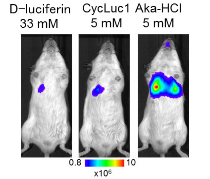
Figure 3. Bioluminescence imaging of lung tumor
Immediate applicability
Owing to its superior properties that enable higher sensitivity and accuracy, AkaLumine-HCl has potential to become the preferred choice for bioluminescence imaging. Nonetheless, for now, the benefits that its discovery brings can already be reaped in bioluminescence imaging studies in small animal models.
Reference
Authors: |
Takahiro Kuchimaru1, Satoshi Iwano2, Masahiro Kiyama2, Shun Mitsumata1, Tetsuya Kadonosono1, Haruki Niwa2, Shojiro Maki2, Shinae Kizaka-Kondoh1* |
Title of original paper: |
A luciferin analog generating near-infrared bioluminescence achieves highly sensitive deep-tissue imaging |
Journal: |
Nature Communications |
DOI : |
School of Life Science and Technology
—Unravel the Complex and Diverse Phenomena of Life—
Information on School of Life Science and Technology inaugurated in April 2016
Further information
Professor Shinae Kizaka-Kondoh
School of Life Science and Technology, Tokyo Institute of Technology
Email skondoh@bio.titech.ac.jp
Tel +81-45-924-5800
Assistant Professor Shojiro Maki
Graduate School of Informatics and Engineering, The University of Electro-Communications
Email s-maki@uec.ac.jp
Tel +81-42-443-5493
Featured News


![]()